The Process of Electroporation
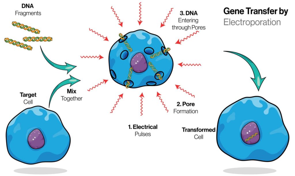
The Steps Of Electroporation

Electroporation is a technique for introducing molecules into cells using an electrical field. Electroporation is often used to introduce DNA or RNA into a cell, but it can also be used to insert protein and small molecules. For example, electroporated DNA can be used as a vaccine or to deliver genes into yeast cells for the expression of proteins that are otherwise not produced by these organisms.

In this blog post, we will discuss the steps involved in the electroporation process along with its applications.

What is produced from electroporation?

Electroporation is an invasiveness-reducing technique for introducing molecules into cells using an electrical field. It is used to introduce DNA, RNA, and siRNA into cells as well as plasmid DNA (in this case, it’s called electrotransformation).

How does electroporation work?

Electroporation is a technique for introducing molecules into cells using an electrical field. The electric field opens the cell membrane to allow the molecules to enter, but only in a tiny area around where the electric field is applied. This will enable you to introduce substances into specific parts of your cells with great precision.

6 Steps to the Electroporation Process

Electroporation is a process that uses a combination of electrical and chemical forces to create temporary pores in the cell membrane. The pores, which are created when an electric field is applied across the cell’s membrane, allow for substances to enter the cell.

Electroporation can be used to introduce DNA, RNA, or siRNA into cells. The procedure uses a mild electric field to create pores in the cell membrane and enable the introduction of molecules.

In the most basic terms, the electroporator device applies an alternating current at a low voltage (< 3000V). The electric field causes the cell membranes to become porous, allowing entry of DNA and other large molecules which otherwise would not pass through the membrane due to their lipid composition and size (about 4 nm).

Here are six steps to the electroporation process:

Collect and prepare the cells you want to transfect.

You should use an aseptic technique when collecting and preparing cells for electroporation. This means that you should avoid contaminating the cells with anything that isn’t sterile. You should also work in a clean, dry environment free of dust or other contaminants so that your cells remain uncontaminated. If you’re working with animal models, it is important to use anesthetics during this process in order to keep animals from experiencing pain or distress.

Sterile materials are critical for the success of your electroporated transfection experiment because they help ensure that everything stays clean and safe for your cells during the collection and preparation steps. You can purchase sterile gloves at most pharmacies or online; however, contact us if you need help acquiring them!

Add the appropriate reagents (e.g., DNA, RNA, siRNA) to the cell solution.

Association of plasmids

Add the appropriate reagents (e.g., DNA, RNA, siRNA) to the cell solution.

DNA and RNA are added to a culture of cells in order to introduce additional genetic material into the cell nucleus. The process is called transfection or transformation. The amount of DNA or RNA used depends on several factors:

  • The type and variant of the target cell line being used; some cells take up more DNA than others do
  • Whether you are using a plasmid or a vector to deliver your gene(s) into the cell
  • What kind of transfection method you will use (e.g., electroporation vs lipofection)

Apply a short electric pulse to your DNA/RNA-cell mixture.

Once you have completed the above steps, there is one final step. You will apply a short electric pulse to your DNA/RNA-cell mixture. It is important that you use the correct parameters for your cell type and voltage level when doing this step.

The best way to determine what these parameters are for your specific situation is to do some research on reputable sources like PubMed or Wikipedia. Once you’ve decided on an appropriate voltage level, length of time, and frequency (or current), plug it all into your electroporation machine and apply it to the sample!

Incubate the cell mixture at 37°C for about 3 hours.

Incubate the cell mixture at 37°C for about 3 hours. Incubation time depends on the cells and the DNA you are using, so it’s best to consult with your instructor or lab manual if you’re unsure of how long to incubate. The incubation should be done in a humidified chamber set at 37°C.

Analyze the gene of interest and measure expression.

The gene of interest is sequenced and the expression level is determined by measuring the abundance of mRNA using qPCR. Fluorescent reporter genes such as GFP allow you to visualize cells and quantify protein levels, while fluorescent markers can be used to track electroporated cells in culture. The marker can also be an enzyme that catalyzes a reaction when it binds a substrate or reacts with another molecule.

We hope that this post has been helpful in understanding the steps involved in the electroporation process. If you would like to learn more about MGMR technology, or how it can be applied in emerging medical treatments., feel free to check out our blog, or contact BioPact

What Are The Types Of Drug Delivery Systems?

Drug delivery systems

Drug delivery is a crucial part of medical care today. The goal is to get the right amount of medication into the bloodstream at the right time and place. It is an important process in the pharmaceutical industry, as it allows for more effective and efficient delivery of medications. There are three main types of drug delivery systems, each with its own advantages and disadvantages.

This article discusses the types of drug delivery systems and how drugs are transported throughout the body.

What are drug delivery systems?

Drug delivery is the method of administering a pharmaceutical compound to achieve a desired therapeutic effect. Drug delivery systems are used to deliver drugs to the patient. Drug delivery systems are used in hospitals, clinics, and pharmacies.

Drug delivery systems are devices used to administer drugs. They can be a pill, liquid injection, or inhaler. Drugs usually enter the body through the skin, mouth, nose or ears. A drug delivery system must deliver its cargo into cells and tissues with minimal loss of potency over time. Drug delivery may also involve other factors, such as product consistency and dispensing method.

Types of Drug Delivery Systems & Their Benefits

Drug delivery systems are designed to deliver drugs, often in a specific way. While all drug delivery systems are meant to get medication into the patient’s body, not all do it in the same way. Drug delivery is an important part of any medication’s treatment regimen because it can help control how much medicine goes into the patient and when they receive it.

There are several different types of drug delivery systems. These include specifically targeted drug delivery systems, modified release drug delivery systems, and combination drug delivery systems. The details of these delivery systems are beyond the scope of this article, but we will focus on some examples and principles related to each type.

There are 4 methods of administering drug delivery systems:

  • Oral (by mouth)
  • Inhalation (through the nose or mouth)
  • Subcutaneous (under the skin)
  • Intravenous (into a vein)

The following are types of drug delivery systems:

Targeted Drug Delivery Systems

Targeted drug delivery systems deliver drugs directly to their target site in the body. This can be done through various mechanisms, including direct injection into the area where you want the medicine (such as your knee joint), oral administration that bypasses digestion by going directly into your bloodstream, or injection into an artery branching off from your heart so that it will flow directly throughout your body. Because these types of targeted drug delivery systems don’t require processing through the digestive system first before reaching their target areas in the body and because they use less medication compared with other forms of administration methods (like pills), they tend to have fewer side effects than other methods.”

Targeted drug delivery systems are designed to either encapsulate the drugs or target a specific area within the body. This is useful in treating patients with conditions such as cancer, ulcers, and inflamed arteries because patients can be given smaller doses of potent drugs over longer periods for more effective treatment.

Benefits of Targeted Drug Delivery Systems

Targeted drug delivery systems offer benefits for patients in many ways. First, they allow the use of smaller doses of potent drugs over longer periods. This means patients can be given the same amount of medication without having to take higher doses, which can cause unpleasant side effects such as nausea or vomiting. Second, targeted drug delivery systems are better at targeting specific areas to treat a disease than traditional methods. They can deliver medication directly to the part of your body affected by the disease, reducing complications and improving your overall health. Finally, some types of targeted drug delivery systems can help reduce or even eliminate pain caused by certain conditions, like cancer or inflamed arteries (a condition called angina).

Modified release drug delivery systems

Modified release drug delivery systems are used to deliver medication over an extended period. This can be beneficial for patients who need consistent, long-term treatment—especially when the body does not easily absorb the medication, or if they have trouble taking their medication daily.

Modified release drug delivery systems are also called controlled release, extended-release, and sustained release. They are designed to control how fast the drug is released into the bloodstream. The most common modified release system is time-release technology. This means the medication is released gradually over time rather than all at once.

Benefits of Modified Release Drug Delivery Systems

There are several benefits to using these types of delivery systems. First, they allow for the slow and steady release of drugs in the body over time, rather than all at once. This means the patient will not experience as many side effects, since there is not as much concentrated dose in one area at one time. It also allows for more consistent dosing throughout the day, since it is released over time instead of all at once.

Combination of Drug Delivery Systems

Combination-release drug delivery systems are a type of drug delivery system that uses two or more drugs to treat a single condition. The most common combination release drug delivery systems are pills and patches, but some examples of other combination release drug delivery systems include pills that contain both an opioid and acetaminophen, a pain reliever; patches that contain nicotine and an antidepressant; and tablets with two different drugs in them.

In these cases, the two drugs are released at different times. For example, a pill that contains both an opioid and acetaminophen is designed so that one drug will be released into your system as soon as you take it (the opioid), while the other drug is released over time (acetaminophen). This allows you to take less of each drug than you might need if they were both released immediately after taking them. It also reduces your risk of experiencing side effects too quickly, because they’re being administered at different times.

Benefits of Combination Release Drug Delivery Systems

One of the advantages of using combination-release drug delivery systems is that they can be tailored to meet your specific needs. If you have a chronic illness, for example, and need to take multiple medications throughout the day, a combination drug delivery system would allow you to take one pill once per day instead of several pills at different times during the day.

Another advantage is that combination-release drug delivery systems are much more effective than traditional oral delivery methods. Oral delivery methods rely on stomach acids to break down the medication before it reaches your bloodstream. This means that some medications may not work effectively, because they don’t get into your bloodstream in time or at all.

There are many types of delivery systems for drugs, the study of which is a very extensive discipline area used in the pharmaceutical development process. However, it is still necessary to develop new technologies that can improve the existing delivery systems. Improving drug delivery systems can help to further optimize the treatment for the disease in question and provide more effective drugs for patients suffering from certain diseases such as cancer.

Understanding The Different Types of Cellular Transport

Different Types of Cellular Transport

Cells need to be able to move substances in and out of their boundaries. This is called cellular transport, and it’s one of the most important functions of living cells. As a result, cells have evolved many different ways to perform this function—different types of cellular transport exist within each type of cell. Here we’ll discuss some common types: active transport, passive transport, osmosis, facilitated diffusion, and vesicle transport.

Active Transport

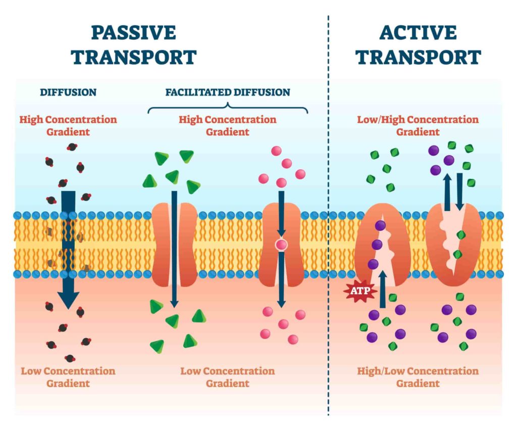
Active transport is a method of transporting substances across a membrane against their concentration gradient. Active transport requires energy, which is provided by ATP. The strength and speed of active transport is proportional to the concentration gradient being crossed so that high concentrations exert stronger forces on the molecules being transported than low concentrations do. Active transport involves the use of proteins called pumps or carriers (depending on what exactly they carry). One example of active transport is the sodium-potassium pump in animal cells, which moves sodium ions out of cells and potassium ions into them.

Passive Transport

Passive transport is the movement of substances through a cell without the use of energy. It is one of two types of cellular transport, along with active transport. Passive transport does not require energy because it does not involve any active process within the cell.

Passive transport occurs in two ways: simple diffusion and facilitated diffusion. Simple diffusion occurs when a substance moves from an area where it’s higher concentration to an area where it’s lower concentration. This movement happens naturally due to differences in concentrations between areas, though no energy is required. Facilitated diffusion involves proteins that help move substances across membranes more efficiently than simple diffusion would allow. This type of passive transport requires energy but is still considered passive because it doesn’t require active processes within the cell itself.

Osmosis

Osmosis is the process by which water moves through a semi-permeable membrane from an area of higher concentration to an area of lower concentration. In osmosis, the water molecules move down a concentration gradient. This occurs especially in plant cells, where water flows into the plant through osmosis. The cell membrane will allow small molecules like oxygen and carbon dioxide to pass through it but not larger substances such as sugar or salt, which would damage the cell if they were allowed inside it.

Facilitated Diffusion

The second group of mechanisms is facilitated diffusion, which uses a carrier molecule to transport substances across the membrane. In this case, the substance moves down its concentration gradient, but with help from a transporter molecule that binds to it and carries it across the membrane.

As an example of facilitated diffusion, consider glucose within a cell: it has a higher concentration outside the cell than inside. But because it can’t pass directly through lipid bilayers, glucose needs to use special carrier proteins called GLUT transporters that act as “doors” into our cells.

Facilitated diffusion has several advantages over other types of cellular transport:

  • It requires less energy than active transport or endocytosis because it does not require ATP for its operation (the energy comes from moving down an electrochemical gradient). This makes facilitated diffusion relatively easy on your body—in fact, many of your organs rely heavily on this type of process to keep working properly!
  • Some viruses (such as HIV) can also use facilitated diffusion to infect cells to reproduce themselves. However, this type of infection usually results in disease, rather than helpful outcomes like those seen during nutrient absorption.

Vesicle Transport 

Vesicle transport is the movement of small packets of fluid (vesicles) through your cell. Vesicles are created by the cell membrane and then filled with water, dissolved gases, or other substances before being transported from one part of the cell to another. The movement of vesicles is powered by ATP (adenosine triphosphate).

Cells need to perform cellular transport to survive.

Cellular transport is one of the most important cellular processes, and cells must perform it to survive. Without this process, nutrients would not enter or leave a cell. Waste could not leave, and information could not move into or out of a cell for protein synthesis or DNA replication.

Without cellular transport:

  • Food would not be able to enter your body’s cells.
  • Waste products would build up inside your body’s cells and eventually kill you.
  • Information from DNA strands (genes) could not be put together into proteins that are essential for life to exist in humans, animals, and plants.4

In summary, while there are several types of cellular transport, they all serve a similar purpose: to move molecules around within cells. Cellular transport is the process that allows cells to move nutrients, hormones, and other molecules into and out of them.

Cells need to perform transport to survive and function. Without cellular transport, you would not be able to move food through your digestive system or oxygen into your lungs. There are several types of cellular transport, each with a specific role. To learn more about cellular transport, and MGMR technology, feel free to check out our blog or contact BioPact

Why Is Cellular Transport Important?

Cellular transport

A cell is a dynamic place, with a steady stream of proteins and other molecules coming in and out. Some proteins are made in the cytosol, but many more are synthesized in the nuclei of cells. To ensure that proteins made in the nuclei can be used appropriately in the rest of the cell, those proteins must be transported out of the nucleus (and mitochondria).

The process through which proteins are transported into and out of cells is known as cellular transport. This blog post will break down several aspects of cellular transport to show you what it is, what it does, and why it is important.

What is cellular transport?

Cellular transport is the process by which substances travel in and out of a cell. This includes the movement of materials like oxygen, nutrients, and waste products through the cell membrane. A substance’s ability to move through a cell depends on its concentration gradient and size relative to other molecules in the cell membrane.

Cellular transport is the movement of materials and molecules within a cell. It’s essential for life and occurs in both plants and animals. Cellular transport can be divided into two groups: passive and active. Passive transport does not require energy to occur; it simply relies on diffusion, where materials move from an area of higher concentration to an area of lower concentration. Active transport requires energy (usually provided by ATP) to push or pull substances against their concentration gradients (the direction in which material will be pushed or pulled by diffusion).

The Importance of Cellular Transport

Cellular transport is the movement of substances and molecules within cells. It is a very important part of cell function because it allows nutrients to be delivered to the cell, it helps remove waste materials from the cell, and it can also maintains homeostasis. Without it, cells would not be able to function properly.

Cellular transport relies on ATP (adenosine triphosphate). This molecule acts as a fuel for many different processes within cells. This process is extremely important for keeping the body healthy and functioning properly. If cellular transport were to stop, then many of these vital processes would cease as well. It’s used by muscle cells for contraction, nerve cells for firing signals, red blood cells for carrying oxygen throughout the body and so much more!

The process of cellular transport is necessary for many different functions in the body, including:

Immune system function

  • The immune system is the body’s defense against infection. It consists of a network of cells, organs, and molecules that work together to protect you from disease. When your body detects an invading pathogen (such as a virus or bacteria), it mounts an attack by sending out specialized proteins called antibodies. These antibodies bind to the invading organism and signal other immune cells to destroy it. The immune system also produces white blood cells that can fight off viruses and bacteria. The lymph nodes, bone marrow, and thymus gland are the main parts of your body’s immune system.

Digestion

  • The breakdown of food into smaller molecules that can be absorbed through the gut wall into the bloodstream. Most of the digestion occurs in the small intestine. The stomach secretes hydrochloric acid and pepsinogen to break down food into smaller particles. After that, the pancreas releases enzymes into the small intestine to further digest food until it is small enough for intestinal cells to absorb it. Absorption Once digested food enters the bloodstream, it must be transported from there to other parts of the body where it can be used as fuel or building blocks for new tissue.

Circulation

  • The movement of blood throughout the body. The heart pumps blood through the arteries, which carry it to the organs and tissues. The blood then returns to the veins, where it flows back to the heart and lungs for oxygenation. Cellular transport is also important for waste removal from the body. Waste products are removed from cells by passing them into a fluid in between cells (interstitial fluid) or by excreting them into urine or stool via exocrine glands.

Oxygenation

  • The transportation of oxygen from the lungs to where it is needed in the body. Cellular transport is also critical for the maintenance of healthy tissues. It’s important that cells get the nutrients they need and remove waste products from their environments. Without proper cellular function, cells would not grow, divide and repair themselves properly.

Excretion

  • The removal of waste products from your body via your kidneys or bladder. The kidneys are the organs that help your body excrete waste products, such as urea and creatinine. The kidneys also filter blood to remove excess water and salts. Excretion is the process of getting rid of unwanted substances in your body through urine or stool.

What is the process of cell transport?

A cell is the structural and functional unit of life. It is the smallest unit of an organism that is classified as a living thing. Without cellular transport, substances would not be distributed throughout your body.

The process of cell transport also referred to as cytoplasmic streaming or fluid flow in the cytoplasm, is a vital function that helps maintain the structure, health, and stability of all cells. The way it works is a little complicated, but it’s not too hard to understand!

There are four steps to the process of cell transport:

  1. A cell’s membrane has special proteins embedded in it that allow it to move by itself.
  2. These proteins get activated when they sense a certain chemical (called ATP). When they do, they pull on the membrane and cause the cell to move.
  3. As this is happening, other proteins in the membrane push against the outer surface of the cell and push it forward.
  4. This whole process repeats as long as there is enough ATP available, and the cell moves away from its original location.

This is a very basic explanation of how cell transport works. It’s easy to get confused by all the technical terms and processes, but it’s important to understand how cells move. This knowledge can help you learn more about cell biology and how to treat certain diseases.

Types of Cellular Transport

Cellular transport is the movement of substances across membranes, and it’s essential for life. Transport proteins move sugars in your body, cells in your heartbeat thanks to ATP-dependent pumps that use ATP as a source of energy, and sodium ions move in and out of cells through ion channels. Without transport proteins, we wouldn’t be able to eat or breathe—or even move!

There are a few different types of cellular transport:

Active Transport

Active transport requires energy to move molecules against their concentration gradients. Examples include endocytosis, where cells take up material outside the cell by enclosing it inside an internal membrane vesicle that then fuses with the cell membrane, and facilitated diffusion, which takes place when specific molecule-protein complexes enable another molecule to pass through membranes more easily than it could otherwise.

Passive Transport

Passive transport does not require energy, because molecules move along a concentration gradient without being altered themselves. Examples include simple diffusion across membranes and osmosis (the net movement of a solvent across a semipermeable membrane).

Osmosis

when water moves from an area with low solute concentration to one with high solute concentration, this is called osmosis. When there are fewer particles dissolved in water than there are in other parts of your body (like blood), osmosis will pull them out of those areas into the solution that has more particles dissolved in it—which means water will move out of your cells and into your blood vessels (this keeps your blood vessels from being too dry).

Diffusion

When molecules move down their concentration gradient (from more concentrated to less concentrated), this is called diffusion. Diffusion is not very efficient, but it’s how most substances get into and out of cells.

Facilitated diffusion

Facilitated diffusion refers to a way that substances move through membranes by using proteins embedded in them called carrier proteins. If there’s an excess of some molecules on one side of a membrane and not enough on the other side, they’ll use these proteins to travel across it and fill up their own side with them again. This process can happen without any chemical energy being used at all (it’s just molecular crowding).

Vesicle Transport

Vesicle transport is a form of diffusion that happens in the cell. It’s used by cells to move substances from one place to another within their structure, like when proteins are sent from the endoplasmic reticulum (ER) to the Golgi apparatus for processing or when lysosomes are sent to the cell membrane so they can break down materials outside it.

Ultimately, cellular transport is an efficient process that helps keep our cells in tip-top shape. IA cell is the structural and functional unit of life. It is the smallest unit of an organism that is classified as a living thing. Without cellular transport, substances would not be distributed throughout your body. t’s seldom talked about (even though it happens on a regular basis), so hopefully this article has given you some insight into this important facet of biology. If you would like to learn more about MGMR technology and its potential within the medical industry, BioPact is here to help. Contact us today. 

What Are Lipid Nanoparticles?

lipid-nanoparticles

Lipid nanotechnology is an area of nanotechnology, which explores methods to modify lipids in order to harness desirable properties that are unavailable with unmodified lipids. Lipids are a broad group of naturally occurring molecules, as well as synthetic entities. It is an interdisciplinary field involving chemistry, biochemistry, molecular biology and physics.

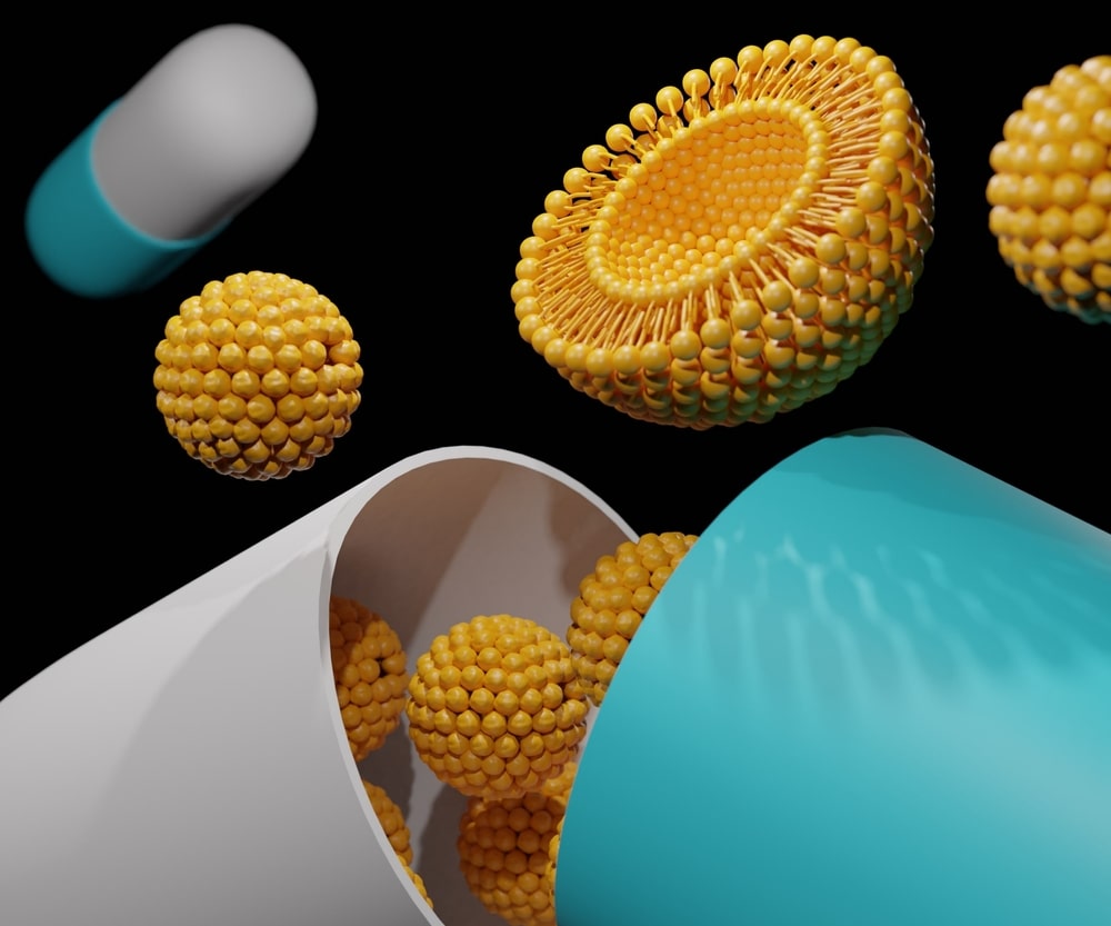
Lipid nanoparticles are tiny capsules of fat and protein molecules, with a diameter just large enough to transport drugs through the body. They’re made from a single type of biomolecule—typically lipid or polyketide—and typically deliver one drug molecule at a time. This simplicity is what makes them so appealing for the drug industry: Lipid nanoparticles can be produced rapidly, cheaply, and efficiently in large quantities.

Lipid Nanoparticles Definition

Lipid nanoparticles are tiny particles composed of lipids, or fats. These types of nanoparticles can be used in a variety of ways including drug delivery, food and cosmetics. They have anti-inflammatory effects and can also prevent infection from bacteria as well as inhibit cancer cell growth.

For example, a lipid nanoparticle could be developed to deliver drugs that target cancer cells. These particles would then be administered intravenously and travel through the bloodstream until they encounter tumor cells. Once they recognize these cells, the lipid nanoparticles would enter them and release their payload.

Lipids are a type of molecule found in both plants and animals. They make up the cell membranes of all living creatures, as well as many bacteria. Lipids are also the building blocks for hormones such as testosterone, estrogen and progesteronLipid nanoparticles are tiny particles composed of lipids, or fats. These types of nanoparticles can be used in a variety of ways including drug delivery, food and cosmetics.

What are lipid nanoparticles made of?

Lipid nanoparticles are made up of lipids, which are a type of fat that is found in many living things. They are composed of long chains of carbon atoms with hydrogen, oxygen, and nitrogen added. The chemical composition determines the shape and functionality of a lipid nanoparticle.

Lipid nanoparticles are typically made of one or more types of lipids, including:

  • Phosphatidylcholine (PC)
  • Phosphatidylethanolamine (PE)
  • Phosphatidylserine (PS)

Lipid nanoparticles are used in a variety of applications including drug delivery, food and cosmetics. They can be made from a variety of materials including soybean oil and fish oil.

How do lipid nanoparticles work?

Lipid nanoparticles are a type of drug delivery system that uses lipids to deliver drugs to specific cells in the body. In the body, lipids (fats) are stored as droplets in the cytoplasm of most cells. The structure of these droplets makes it possible for lipid nanoparticles to carry drugs and infiltrate specific cells by carrying drugs that can be absorbed by the cells in question.

Advantages of Lipid Nanoparticles

There are a number of advantages to using lipid nanoparticles.

Targeted delivery of medications

One of the most important advantages is that they can be used to target specific cells. This means that they can deliver drugs and other substances directly to a specific cancer cell, for example, and only affect those cells. This makes them very useful in medicine and other areas where targeted delivery is important.

Stability

Lipid nanoparticles are more stable than liposomes. This is because lipid nanoparticles are made out of synthetic or natural polymers, which do not have the same tendency to degrade as liposomes do. This means that the drug will remain intact for longer in your body and be able to reach its target cells at a higher percentage rate than other types of delivery systems can.

Increased bioavailability of drugs

Lipid nanoparticles have been shown to increase the bioavailability of drugs by creating a bio-lipidic barrier on their surface, which protects them from degradation when they enter into your bloodstream. In addition, this layer allows them to penetrate through cell membranes much more easily than other delivery strategies can because there’s less resistance when they come into contact with these surfaces (since they’re mostly made up of phospholipids).

Easily modified

Lipid nanoparticles can be easily modified to encapsulate a wide range of drugs and other substances, making them versatile in terms of what they can be used for. They can be modified to change their physical characteristics, such as size and surface charge. Depending on what you want them to do, they can also be engineered to have different shapes (e.g. spherical or fibrous).

Are lipid nanoparticles safe?

Lipid nanoparticles are safe for use in humans, animals, plants and the environment. Lipid nanoparticles can be used to deliver drugs to targeted tissues or organs and can be adapted for oral or intravenous delivery of drugs. The unique properties of lipid nanoparticles make them well suited to treat a variety of diseases including cancer, neurological disorders and cardiovascular diseases.

Due to their small size, lipid nanoparticles are able to penetrate the blood-brain barrier and other cellular barriers in the body. This makes them an effective delivery system for treating diseases that affect specific organs or tissues.

Lipid nanoparticles are a promising treatment for cancer, but they are not a silver bullet. To determine the best way to use them in a particular patient, doctors will need to consider factors like the type and stage of cancer being treated, as well as other treatments available.

What is Transfection & How Does It Work?

transfection definition and uses-min

Transfection is the introduction of genes or other DNA into cells by a non-mechanical method that does not involve electroporation. Transfection can be carried out with a variety of delivery methods.

The goal of transfection is to introduce genetic material into a cell so that it can be expressed within the cell. This can be done for a variety of purposes, including in vivo gene therapy or research applications.

Transfection Definition

Transfection is a process that involves the introduction of foreign DNA into a cell to produce genetically modified cells. The process involves the use of a vector (a virus or a plasmid) to introduce DNA into the host cell. The vector is then able to reproduce itself and spread its genetic material throughout the host cell until it reaches every part of it. This can take place either inside or outside of an organism’s body, depending on what kind of experiment you’re performing.

To understand transfection, you first need to know about gene therapy and how it works. Gene therapy involves replacing or modifying defective genes with healthy ones. In gene therapy, researchers use viruses to deliver healthy genes into the cell’s nucleus. This is called viral transduction because the virus has been modified so that it will only infect certain types of cells. The virus then releases its payload of healthy genes inside the cell where they can integrate into its DNA and replace any defective genes it finds there.

This can be done by either transient or stable transfection.

  • Transient transfection results in only temporary changes in the host cell’s genetic makeup, and it is often used to study gene expression or protein production.
  • Stable transfection involves the introduction of permanent alterations in the genome of a host cell, which causes it to permanently express new proteins or genes.

Transfection is commonly used to introduce genes into cells for research purposes, but it can also be used to treat diseases. Transfection can be accomplished using liposomes or other carriers that contain DNA molecules. These carriers are typically constructed from lipids or other small molecules that interact directly with lipids on the cell surface.

When performing viral transduction on cells that are not dividing (like neurons), researchers use liposomes (tiny spheres made of lipids) instead of viruses as delivery vehicles because they are easier to control and less likely to cause side effects than viruses would be. Liposomes contain molecules that allow them to fuse with a cell’s membrane and release their cargo inside

What is the purpose of transfection?

Transfection is often used to introduce genes into cells and tissues, in order to study the effects of these genes or to produce specific proteins through protein production.

A common use for transfection is gene therapy, which involves using transfection to insert a healthy copy of a mutated or missing gene into a patient’s cells. Transfection can also be used to deliver corrective genes when treating genetic disorders, such as sickle-cell disease or cystic fibrosis.

Researchers have developed several methods that allow them to increase the efficiency of transfection and reduce toxicity levels associated with this method. These include electroporation (electrical stimulation), lipid-based delivery systems like liposomes and cationic polymers, viral vectors such as lentiviruses, adenoviruses, and retroviruses (DNA viruses), as well as mechanical methods like pressure injection.

How does transfection work?

Transfection is a process in which a gene, or set of genes, is introduced into cells. It’s used to manipulate the genetic material of cells and can be used to treat various diseases or disorders.

It’s done with a vector, which is a piece of DNA altered so that the cell can recognize it as its own genetic material. The vector is usually placed inside an empty virus particle, or “virus-like particle” (VLPs), which injects the genes into the cells.

The goal of transfection is to get your desired genetic material inside cells so that they can begin expressing that material and producing proteins from it. This can be done by using viruses or plasmids as vectors for your DNA—both are capable of carrying large quantities of genetic information from one place to another. Viruses enter cells through their membranes and fuse with their cytoplasmic membranes, releasing their genetic material into their new host’s cytoplasm where they are able to replicate themselves exponentially until they have taken over all available space within those cells (which eventually leads them to burst out in order to release more copies).

Transfection uses viruses as vectors because they’re incredibly effective at getting their genetic material inside cells—and they can’t replicate themselves once they’ve done so. That means there’s no risk of the infected cell becoming cancerous through viral replication, unlike other methods of gene therapy that introduce foreign genes directly into cells (such as when scientists create modified stem cells).

Types of Transfection

Transfection is the process of delivering genetic material into cells. There are several ways to do this, and in this article we’ll discuss five common methods:

Lipofection

Lipofection utilizes lipid vesicles to transport DNA or RNA into cells. These lipid vesicles are formed by mixing DNA, cholesterol, and other components in an oil-in-water emulsion. When this mixture is added to a solution containing cells, it creates tiny bubbles that surround the cells and allows them to pull in the contents of these bubbles and become transfected with your genetic material.

Electroporation

Electroporation uses an electrical current to create holes in cell membranes; this causes foreign protein molecules and other substances within these holes (including DNA) to enter the nucleus of a cell where they can be integrated into its genetic structure.

Biolistic bombardment

Biolistic bombardment (also known as particle bombardment) uses microscopic gold particles coated with DNA to create holes in plant or animal cells. The gold particles are shot through a gun at the cells, which are then hit with a puff of air that causes them to open their membranes, allowing DNA molecules to enter.

Gene transfer

Gene transfer is most often used to create genetically modified organisms or GMOs. These are plants, animals, and bacteria that have been altered at the genetic level so they can perform certain functions better than their non-modified counterparts.

Cell fusion

Cell fusion uses two cells of the same type that fuse together and form one new cell. The process is often used in creating genetically modified organisms, or GMOs. The process is also used to create hybridomas, which are cells that grow immune to cancer and viruses.

Nuclear transfer

Nuclear transfer can be used in research or medical applications. It involves removing the nucleus of a cell—which contains DNA—and transplanting it into an empty egg cell that has had its own nucleus removed. When this new cell is implanted into a woman’s uterus, it forms a fetus.

Benefits of Transfection

The benefits of transfection include:

Reduction in cell death

Transfection can protect cells from apoptosis, or cell death. This is because transfection can deliver genes that will prevent apoptosis, as well as other harmful effects of gene expression. -Increased research: Transfection allows researchers to study specific diseases and their causes in more detail. This process allows scientists to better understand what causes a disease, how it progresses over time, and how it might be treated.

Improved protein production

Transfection can increase protein production in cells. This is important because proteins are the primary components of cells. They perform many functions, including transport of nutrients, cell growth and division.

Improved cell growth and survival

Transfection can increase cell growth and survival rates. This is important because it allows researchers to study how cells react to certain stimuli, such as changes in pH or temperature. These studies can lead to a better understanding of the effects of disease on cells and help scientists develop new treatments for various conditions.

Improved gene expression

The introduction of a gene into a cell can lead to improved expression of that gene. This is important because it allows researchers to study how cells react to certain stimuli, such as changes in pH or temperature. These studies can lead to a better understanding of the effects of disease on cells and help scientists develop new treatments for various conditions.

Biopact Transfection Technology

Biopact Transfection Technology is a method of introducing genes into cells. Biopact Transfection Technology uses a synthetic, non-viral carrier called PTX1 that binds to DNA and carries it into living cells. The technology has been shown to deliver many types of DNA molecules, including plasmids, viral vectors and siRNA.

Biopact Transfection Technology can be used for gene therapy applications and basic research studies related to genetic engineering or gene editing. This method allows you to introduce your genes into almost any kind of cell line (stem cells included). It’s important because it gives scientists access to previously impossible levels of cellular modification with their own selected genes or RNAi sequence, without using viruses as carriers!

Electroporation: Definition, Uses, & Benefits

electroporation-definition

Electroporation is a method of transfection used to facilitate the transfer of DNA, RNA, proteins, and small chemical molecules into cells. Electroporation can also be referred to as electropermeabilization. This method is used in large-scale protein production and even cancer therapy.

What is electroporation?

Electroporation is a technique used to introduce DNA, RNA, proteins, and small chemical molecules into cells. It is particularly useful for large-scale protein production and even in cancer therapy. This technique can be used on bacteria and yeast because it does not require the use of transformation or transfection agents.

How does electroporation work?

Electroporation is a method of introducing DNA into a cell. It uses a short electric pulse to create pores in the cell membrane. This allows the DNA to enter the cell and become part of its genetic makeup. The pores are temporary and close after a short period of time, so they won’t allow toxins or other harmful substances to enter your cells.

Electroporation is a technique used to facilitate the transfer of DNA and other molecules into a cell. When cells are exposed to an electric field, their membranes become permeable and can be subsequently fused by various agents such as DNA or lipids.

What is electroporation used for?

Electroporation is used in:

Genetic engineering

The most common use of electroporation is in genetic engineering. A small electric pulse is applied to cells containing a piece of DNA that you want to transfer into the cell’s genome. This allows some of the DNA to enter the cell and become part of its genetic makeup. The pores are temporary and close after a short period of time, so they won’t allow toxins or other harmful substances to enter your cells.

Protein production

Electroporation is used to produce proteins in bacterial cells. This can be done by inserting a gene that codes for the protein of your choice into the genome of a bacterial cell. The electric pulse opens up pores in the cell’s membrane, allowing messenger RNA (mRNA) molecules containing the gene to enter and start producing protein.

Cancer therapy

Electroporation is used to treat cancer by delivering DNA into tumor cells. A virus or other vector carrying the DNA is used as a carrier; it will infect and enter the tumor cell, then release its load of DNA through pores in the cell’s membrane.

Bacterial and yeast studies

Bacterial and yeast studies have shown that electroporation can be used to carry out gene knockouts and other genetic manipulations in bacteria and yeast. This method is used to study the effects of gene knockout or overexpression in a particular organism. It can also be used for large-scale mutagenesis studies, producing hundreds of thousands of strains per experiment.

Creating transgenic animals

Transgenic animals are animals whose DNA has been altered by the introduction of foreign genes. The most common method of creating transgenic animals is to use electroporation to transfer a plasmid containing a gene into embryonic stem cells (ESCs). These ESCs can then be used to produce cloned offspring that carry the new gene.

Creating transgenic plants

Transgenic plants are created by introducing foreign DNA into a plant’s cells. This is usually done to introduce new traits or to silence genes that cause disease. The most common way to create transgenic plants is through the use of Agrobacterium tumefaciens (AT) bacteria. AT bacteria naturally colonize the roots of many plant species, including tomatoes and potatoes, where they can transfer their own DNA into the host genome.

Advantages of Electroporation

Electroporation advantages

It’s a non-invasive

It’s a non-invasive technique that doesn’t require any surgery, incisions or stitches. Instead, it uses an electric field to deliver DNA into your cells by punching holes in their membranes.It is non-viral, non-toxic, and can be used on all cell types including human and animal cells.

It’s non-destructive

Electroporation doesn’t kill your cells or damage their DNA like other methods do. This makes it ideal for studying single cells from human tissue samples—you don’t have to worry about damaging the very thing you’re trying to study!

It’s quick

Electroporation takes only about 30 seconds per sample—much faster than other methods for delivering DNA into living cells like transfection (about 3 hours) or chemical conjugation (1 hour). That means researchers can get back on track quickly after each electroporation experiment and start analyzing results sooner!

Disadvantages of Electroporation

While electroporation is powerful, it does have its drawbacks. As a biological technique, it has limitations that make it unsuitable for certain applications. In fact, there are several situations where electroporation should not be used at all. For example:

Electroporation isn’t always efficient.

Electroporation isn’t always the most efficient method for introducing DNA into cells. The process of introducing DNA into a cell can sometimes be improved through techniques such as transfection (introducing DNA with a virus) or lipofection (introducing DNA with liposomes). Electroporation may also be less effective than these methods because it requires large amounts of energy to open pores in the membrane of biologic structures like cells. This requires more time and voltage than other methods.

Can’t be used in all cells.

Electroporation works on cells with high transfection efficiency, which means they are easy to permeabilize and therefore easily transfected. If you want to use electroporation on a cell line that’s not known for its transfection efficiency, it’s possible you’ll need to do other methods of transfection first in order for the cells to be permeable enough for electroporation.

Can’t be used if you’re using small plasmids.

Because electroporation uses electric fields, which can cause heating and shearing of your DNA, it’s not ideal if you’re using a small plasmid with low transfection efficiency. The smaller the plasmid is, the more likely it will be damaged by the electric field during electroporation.

The technique has been used for decades and continues to be an effective way of transferring DNA into cells. If you’d like to learn more about the potential uses for MDMR gene editing, feel free to view our blog or resources page. 

Gene Editing 101 Guide: What is Gene Editing?

Gene-editing-101

What is Gene Editing? And how does it work? These are questions we need to be answered before we begin this exciting new science. In this article, you will learn what gene editing is, what can it do, and what the ethical and policy issues are. There is no right or wrong answer when it comes to gene editing, but we can start with a fundamental understanding of the technology. Hopefully, this information will help you make an informed decision.

You may be wondering what gene editing is. This complex process involves modifying DNA in the human body by making small, controlled changes. According to the Purdue University College of Agriculture, gene editing is “a scientific innovation”. Dr. William M Muir, a biotechnologist and professor at Purdue, compared gene editing to editing the human genome to rearranging words, punctuation, or paragraphs.

A gene is a series of letters in DNA that give instructions to build a protein. Every cell needs DNA for the same reasons that the human body does. But DNA breaks are not irreparable, and our cells have a mechanism to repair damage to our DNA. To fix a genetic mutation in a person’s body, gene editing uses the cell’s own DNA repair machinery. In cystic fibrosis, for example, a mutation in the CFTR gene makes a person susceptible to the disease.

There are many different types of genome editing. CRISPR Cas9 uses two core components to make edits to DNA. The Cas enzyme cuts DNA and makes edits at a specific location defined by a guide RNA. During the process, the desired changes become permanent. This process is also known as gene therapy. It isn’t yet known if it has any negative side effects.

What is gene editing?

The scientific study of genes and heredity dates back to the mid to late 19th century. In recent years, understanding genes have gained much greater public attention, mainly through the Human Genome Project, a collaborative project that began in the early 1990s to map the human genome. The technology has made it possible to modify and correct harmful mutations and alter the activity of specific genes in plants and animals. But how does gene editing work? Let’s look at some of the potential uses of gene editing in humans.

Today, genome editing has applications in a variety of fields, including medical research and agriculture. Using CRISPR to edit genes, scientists can create knockout models of genetic diseases in animals and study the underlying causes of those diseases. They can also change genes in particular tissues to treat or cure diseases. Scientists have even used gene editing to modify pigs to render them resistant to certain viruses. In the future, pig organs could serve as replacement organs for humans. Various research projects are also using genome editing to modify yeast cells, produce biofuels, and improve strains of agricultural crops.

There are many ways to edit genes, but the latest technique is CRISPR-Cas9, which is more expensive and more complicated. In addition, it can also be applied to animals, allowing scientists to produce healthier food options. These techniques use RNA-guided enzymes called endonucleases, which sever specific DNA proteins on chromosomes. In this way, changes can be made at DNA breakpoints, changing an organism’s genetic makeup.

What can gene editing do?

The idea of gene editing the human germline is controversial and is yet to gain widespread approval. Because it would alter the genes in sperm, eggs, and early embryos, the process would also affect descendants. It could alter human heredity in dramatic ways. For example, hundreds of genes are involved in many common diseases. Editing one gene would decrease the risk of developing a disease and may even prevent it. But, the question remains: what is the best use of gene editing?

Gene editing is a form of DNA repair. Genes contain instructions that tell a cell how to build proteins. The repair of DNA damage is important for the health of cells, and this technique uses the cell’s own DNA repair machinery to fix a person’s genes. Gene editing is an important tool in cancer research because it allows researchers to correct mutations in a person’s DNA. One example of a mutation is CFTR, which is responsible for cystic fibrosis.

However, it’s a major challenge to get the molecules into a human cell. For this reason, scientists are currently testing gene editing in cells and animals. It’s unlikely to be widely available until the technology is safe enough for human trials. In the meantime, gene editing has made strides in cancer research. If successful, it will revolutionize the way that cancer researchers treat patients. It will be easier than ever before to cure a disease using this technology.

How does gene editing work?

Scientists have developed a method known as CRISPR/Cas9 to edit a DNA sequence. The Cas9 enzyme snips a portion of DNA, bringing the attention of the cell’s DNA repair machinery to the break. This process permanently corrects the mutation. It can repair a single gene or an entire group of similar mutations. In this way, it is possible to alter a gene’s function or even turn it on or off.

Although CRISPR is relatively new, it has already shown promise. Developed for use in agriculture, it can improve plant genetic codes. Humans have been breeding plants for thousands of years. CRISPR technology is the most advanced plant breeding tool yet, and it promises to produce stronger and healthier plants. Ultimately, these improved crops will have many benefits for farmers, consumers, and the environment. However, there are still some concerns surrounding the technique.

CRISPR/Cas9 genome-editing techniques have been used in laboratory experiments to cure single-gene disorders. The TALENs genome editing technology has also been used to treat Layla’s disease. It saved her life. Researchers are excited about the potential of CRISPR-Cas9 in battling cancer, ALS, and other genetic diseases. But it is not until it can be used to cure people that the technique has its limits.

Pros and Cons of Gene Editing

The Pros and Cons of gene editing are well documented, but the debates around this technology have been even more contentious. Some scientists question whether the benefits far outweigh the risks, and some have raised concerns that it will exacerbate current problems such as global food shortages and rising prices. However, others cite the need for more research and data before concluding that gene editing is the way to go. 

Pros:

Can help create a treatment for genetic disorders.

Gene editing can help cure people who have genetic disorders or make them less likely to develop such disorders. For example, gene editing could be used to treat sickle cell anemia or Huntington’s disease. Gene editing could also help prevent people from developing certain cancers by changing their genes so that they don’t produce cancerous cells.

Can help create hardier crops

Gene editing could also make crops more resistant to diseases or pests. This would allow farmers to use less pesticides on their crops, which would be better for the environment and human health. It might also result in higher crop yields (more food per acre of land), which would help feed more people worldwide than we already do today!

Offers new possibilities for treating diseases

Gene editing offers new possibilities for treating diseases like Alzheimer’s or Parkinson’s that are currently untreatable or hard to treat with traditional methods such as drugs or surgery; however, it might not work as well as these other treatments once they become available.

Cons:

Cost prohibitive

Gene editing is expensive, and it is currently difficult for researchers to get funding for experiments that involve CRISPR-Cas9 (the most common gene editing tool). This means that many researchers who want to study this technology cannot do so because they do not have enough money to fund their research projects.

Ethical Concerns

There are concerns that the technology could be used for unethical purposes. For example, it might be possible for someone to use CRISPR to make a baby with certain desirable traits, such as being more intelligent or athletic than other people. This raises questions about how we should regulate gene editing and who has access to it.

Side effects are not fully understood

Gene editing is still in its infancy, and there are many unknowns about the potential side effects of gene editing. For example, we don’t know how it will affect the long-term health of the population or if it will cause any genetic problems for future generations.

MGMR Gene Editing

 The MGMR Gene Editing technology is a novel gene editing technology that works by modifying the DNA of T cells to treat cancer. 

In order to use this treatment, doctors first remove T cells from the patient’s blood. They then use a special enzyme to cut out part of the T cell’s DNA and replace it with new genetic material. The modified cells are then returned to the patient’s body, where they grow into mature T cells and begin attacking cancer cells throughout the body.

The MGMR Gene Editing technology was developed by BioPactCT Inc., which has been conducting clinical trials since 2017. In these trials, patients have been treated with modified T cells that target cancerous B cell populations in their bone marrow or other tissues. Results from these trials have shown that the treatment is safe and effective at eliminating cancer cells in patients’ bodies while leaving healthy tissue intact.

What Are Liposomes?

What Are Liposomes?

You may have heard of the term liposome, but what is it and how do they work? This article will explain what liposomes are, what they are made of, and how liposomal technology works. To understand the concept of liposomes, read the following information. 

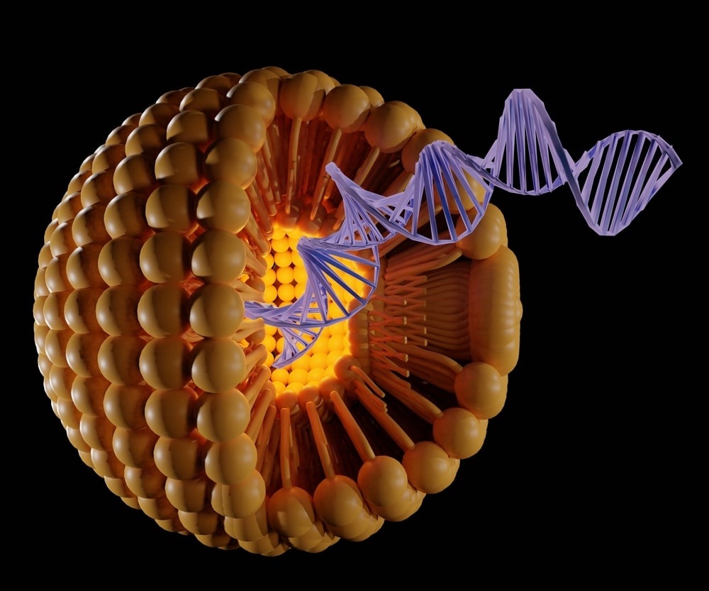
What Are Liposomes? Liposomes are spherical vesicles that contain at least one lipid bilayer. Liposomes are useful as drug delivery vehicles and are widely used in biomedical applications, including the delivery of pharmaceutical drugs and nutrients. In addition, lipid nanoparticles are used in DNA and mRNA vaccines. 

There are a variety of methods for preparing liposomes, with each method having different advantages and disadvantages. The most common method involves dissolving lipids in organic solvents and drying the resultant solution. The dried lipids are then hydrated and isolated, and the liposomes are ready for use. The advantages and disadvantages of each method are discussed in the following paragraphs. Several studies have shown that liposomes have superior effects in treating various diseases.

Liposomes are intracellular vesicles that transport substances into and out of cells. Liposomes are made from phospholipids, a type of fat that makes up cell membranes. They are also found in eggs and are responsible for transporting natural substances. When formed, liposomes contain a water-loving head and two water-fearing tails. The head of the liposome attaches to the acceptee cell’s cell membrane.

When liposomes are filled with nutrients, they are more effective than non-liposomal delivery. They contain phospholipids, which are released by the liver. Liposomes are then passed through the intestines, blood, and body, and enter cells. As a result, liposomes enhance intracellular delivery by more than 100-fold. When a nutrient is encapsulated in liposomes, it travels to the cells.

Liposomes Definition

Liposomal particles are spherical, self-closed structures composed of lipid bilayers and an aqueous compartment. They can consist of one or multiple concentric membranes and may be as small as 20 nm or as large as dozens of um. The structure of liposomes helps them trap a solvent inside. They are also used in contrast-enhanced ultrasound.

Listed below are the three main types of liposomes. These types are used to deliver substances to various organs.

  • MLV (Multilamellar vesicles)
  • SUV (Small unilamellar vesicles)
  • LUV (Large unilamellar vesicles)

A liposome is a small, oblong-shaped particle with a low density of hydrogen. Liposomes differ from other types of particles in two main ways – size and homogeneity. The latter is essential for predicting their stability in solution. In addition to size, the liposomal formulation must have low PDI, which means that a particle’s density is uniformly distributed.

Another major difference between liposomes and vesicles is the polar end of a liposome is asymmetric, so hydrophilic solutes cannot pass through it. In contrast, hydrophobic molecules can bind to hydrophobic molecules that will help deliver their contents. In addition, liposomes can fuse with other substances to increase their ability to deliver drugs. Liposomes are extremely useful for various biotechnology applications.

This study describes a methodology for developing liposomal products. It describes how to use QbD to assess the quality of liposomal formulations. It also discusses the physical properties of liposomes, such as hydration, hydrophilicity, and steric stability. The most important impact area for liposomal products is cancer therapy, which has made liposomes a valuable tool in the treatment of disease.  

What are liposomes made of?

Whether you’re looking for an effective way to deliver life-saving medical treatments or a convenient delivery system for vitamins and nutrients, liposomes are a good choice. These spherical bubbles contain essential fatty acids and phosphatidylcholine molecules. Unlike other encapsulated supplements, liposomes protect your nutrition from the body’s immune system. What’s more, liposomes are able to pass through the gut lining.

Liposomes are widely used in the pharmaceutical and cosmetics industries. Encapsulation by liposomes has been extensively studied in the food industry, as they are a good carrier for hydrophilic and hydrophobic compounds. Unlike other encapsulated compounds, liposomes are able to retain and release the compound they contain. And because they can retain various chemical and physical properties, liposomes are able to transport numerous molecules.

Liposomes are an efficient delivery system for drugs. They encapsulate a portion of an aqueous solution inside a hydrophobic membrane. While hydrophilic molecules cannot pass through liposomes, hydrophobic chemicals can pass through. Moreover, liposomes are versatile and can fuse with other bilayers to deliver the contents. Drug delivery by liposomes is possible through genetic engineering and the encapsulation of drugs.

The discovery of liposomes was first reported in the early 1960s and has gained wide recognition as an efficient drug and cosmeceutical delivery system. Liposomes vary in size and charge depending on manufacturing protocol. Phospholipids are a major component of liposomes and are a safe and effective way to deliver ingredients through the skin and the gastrointestinal tract. And they can be used in various diagnostic procedures, including cancer diagnosis and prevention.

How does liposomal technology work?

Liposomal technology is a form of nanotechnology that encapsulates drugs. It allows them to be delivered to specific locations of the body, where they can maximize the release rate. Liposomes are a good option for the treatment of dietary deficiencies. In addition to its use in nutritional supplementation, liposomal technology may be applied to other conditions.

A lipid bilayer forms the core of a liposome, and its hydrophobic membrane encapsulates an aqueous phase. The lipid bilayer allows the drug to be incorporated into two materials with different solubilities. Cholecalciferol/vitamin D3 is a lipid-soluble substance, while glutathione is a water-soluble compound.

Regular supplements are easily broken down in the stomach. A digestive tract is a busy place, and the enzymes and acids break them down into small fractions. Some nutrients struggle to enter the bloodstream, while others are completely destroyed. Liposomal supplements are a relatively new development, but are widely available in many health food stores. Liposomal delivery of medication is the best option available and may help prevent disease. Antioxidants, B-vitamins, and immune boosters can now be delivered to the target site. The delivery of these nutrients is important for the human body because natural antioxidants are unstable and their bioavailability decreases when dispersed to the target site. If you are prone to intestinal discomfort, liposomal technology could be the answer.

Why are liposomes used in drug delivery?

Liposomal drug delivery is possible because the drugs are encapsulated in liposomes. These encapsulated particles have the ability to reduce drug toxicity and protect the drugs from degradation. These encapsulated liposomes also increase the maximum concentration of drugs in the blood and shorten the time needed to reach the maximum concentration. These properties of liposomes make them suitable for a variety of drug applications.

Liposomes have a unique vesicular structure and contain a lipid bilayer that surrounds an aqueous phase. These vesicles can contain up to 85% of the drug substance. Moreover, they are made from free fatty acids, cholesterol, or polyoxyethylene. They have two to seven bilayer shells with a large amorphous core.

Liposomes are composed of different lipids, but phosphatidylcholine is the most widely used. This lipid is neutral and relatively inexpensive, and it also carries several important functions. Liposomes are generally sterile injectables. They can release drugs either in extracellular or intracellular environments. This allows them to reach their intended location without compromising efficacy.

A number of common applications of liposomes have been discovered. These include antineoplastic treatment, leishmaniasis treatment, and biotechnology. Eventually, they could become a cheaper alternative to lipid formulations used in conventional pharmaceuticals. Some liposomal preparations are already available, such as anticancer, antifungal, and aerosol forms. This makes them a versatile and flexible nano delivery system.

Benefits of Liposomes

Lipid vesicles can protect sensitive ingredients in food products. 

This is mainly due to the fact that liposomes have the ability to pass intact through the stomach and intestine. This means that liposomes can enhance the biological availability of oral active substances. Here are some of the benefits of liposomes for food and medicine. If you want to learn more about this technology, keep reading.

The development of liposomes in the area of neurology has been a focus for scientists

They have recently outlined some of their accomplishments in liposome-based drug delivery across the blood-brain barrier. For the most part, the authors have focused on imaging drug accumulation and glioma diagnosis. In addition, the presence of targeting ligands on the liposomes’ surface has also improved their uptake by cancer cells.

Liposomes are particularly useful for the delivery of hydrophobic compounds. 

These bioactive compounds have very poor solubility in water. These compounds must overcome several obstacles to achieve their bioavailability. Liposomes are particularly useful for hydrophobic compounds, which are poorly soluble in water. As a result, they must overcome their low solubility in water to reach their target organs. Furthermore, liposomes improve the permeability of the GI tract wall, which helps them avoid deactivation in the liver.

Liposomes are the ideal drug delivery system. 

Because of their ability to enhance the sensitivity, specificity, and durability of anti-malignant agents, liposomes are an excellent choice for drug delivery. They also have numerous clinical uses, such as for cancer therapy. The development of liposomes in this area is only just beginning. However, the benefits of liposomes are significant and worth exploring further. The future of cancer treatment rests on the continued development of these drugs and their delivery.

Nanoparticles in Medicine: What Are They & Are They Safe?

Nanoparticles in Medicine

Nanoparticles are becoming more and more prominent in the development of medicine, being used as diagnostic agents, targeted drug delivery vehicles, and novel carriers for oncology treatments.

If you have ever wondered what nanoparticles are, this article is for you. Are nanoparticles safe for the body? How do they work? And what are the benefits and risks of these materials? Nanoparticles in medicine are being used more, but there’s still some controversy over whether they’re beneficial or harmful. Read on to learn more.

Nanoparticles in medicine can deliver medicines to specific parts of the body. These tiny vehicles can be designed to attach to specific cells or organs and transport the medicine directly to those cells. Unfortunately, there are many barriers in the human body that make it difficult for medicines to get to the correct place. In order to overcome these barriers, scientists have developed methods to attach nanoparticles to specific proteins on the cell membrane.

The use of nanoparticles in medicine has many benefits. Their size allows for the development of advanced diagnostics, therapies, and more. In addition to their many medical applications, nanoparticles also present unique environmental and societal challenges. Although nanoparticles can help with many aspects of modern medicine, some research has been conducted on their toxicity and safety. As such, it is important to understand what these particles are and how they can improve human health.

How nanoparticles are used in medicine?

Nanoparticles are small particles with many uses in medicine. Nanoparticles can carry antibodies, drugs, imaging agents, or other substances. These tiny particles are currently being studied for use in cancer diagnosis, treatment, and detection. Ultimately, nanoparticles will help physicians, pharmaceutical and biotech companies better understand the treatment of diseases and improve patients’ quality of life. One of the most widely-used applications of nanoparticles is drug delivery. Numerous studies have shown that these particles can deliver drugs effectively. Most polymer and liposome-based systems are in clinical use today. But while these systems are useful, they also pose unique societal and environmental challenges.. The question is: Which are the best applications for nanoparticles in medicine?

Are nanoparticles safe in medicine?

Nanoparticles are incredibly small particles with the ability to cross cell membranes and reach various organs at a cellular level, including the blood. Nanoparticles also have higher surface-to-volume ratios than larger particles, meaning there are more molecules per square millimeter. While some nanoparticles are safer than others, they still pose unique societal and environmental challenges. As such, the question is, are nanoparticles safe for use in medicine?

There are several aspects to consider when determining whether nanoparticles are safe to use in medicine. While nanoscale materials may appear identical in composition, their size, shape, and surface charge may affect their cellular fate, accumulation in the body, and biological response. To further investigate these issues, researchers must understand the underlying mechanisms of nanomaterials in order to ensure that they remain safe. 

What do nanoparticles do to the body?

Once inside the body, nanoparticles encounter a series of barriers. They encounter the gastric and intestinal milieu, mucus, and tight junctions that block paracellular passage. Nanoparticles also encounter enterocytes, the cells that line the gastrointestinal tract. They may also pass through spaces between cells and enterocytes, but only if they have an antibody to attach to them.

Among the potential hazards of nanoparticles is their potential for accidental inhalation. This is a major concern because many cosmetics, cleaning products, and other products contain nanoparticles. These particles are most likely to accumulate in the nasopharynx, while those with a size between one and five um accumulate in the tracheobronchial area. Approximately one-fifth of nanoparticles are transported into blood circulation.

Benefits of Nanoparticles in Medicine

Nanoparticles are tiny particles that can be used in medicine to deliver drugs and other substances directly to the target site. They have a variety of benefits and can be used to treat a range of conditions, including cancer.

Nanoparticles are useful in several areas of medicine.

 In ophthalmology, for example, they can enhance drug delivery. Unlike oral medications, drug solutions in the eye are usually highly concentrated and must be applied frequently to ensure absorption. Because mucus moves during blinking, it can make the drug solution ineffective if it remains in the eye too long. Therefore, nanoparticle-based drug delivery systems can prolong the duration of residence in the eye.

They can improve vascular function. 

Antioxidant properties of nanoparticles may also improve vascular dysfunction associated with diabetes, atherosclerosis, and hypertension. However, further studies are needed to determine the effectiveness of nanoparticles in these conditions. The benefits of nanoparticles in medicine continue to grow as researchers seek to discover their potential and minimize the potential risks and side effects. But until then, we can take comfort from this fact.

Flexibility 

Nanoparticles can be made from a wide variety of materials, including metals, ceramics, semiconductors, plastics, and polymers. Because they are so small, they are able to cross cell membranes more easily than larger particles and accumulate at the desired location without being filtered out by the kidneys or spleen.

Lower Toxicity Levels

Nanoparticles’ small size also makes them easier for cells to uptake and process than larger particles would. This means that less material is needed for treatment, which leads to lower toxicity levels than existing treatments like chemotherapy or radiotherapy would cause if used alone.